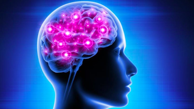
One of the many ways that COVID-19 leaves its long-term impact on patients is on the brain. Since the beginning of the pandemic, scientists have been trying to decipher how exactly the disease and the coronavirus can affect the brain. Earlier, it was thought that coronavirus can enter the brain and infect neurons, the brain cells. However, late researches have shown that this may not be the case.
Nevertheless, evidences are getting clearer about how the brain gets affected and these suggest that the effects can be multipronged. The coronavirus might attack certain types of brain cells directly or reduce blood flow to the brain tissue or trigger immune response that can eventually harm the brain.
A survey published in May this year revealed that 80% of the patients surveyed, who were also hospitalised with COVID-19, developed neurological symptoms. The survey was conducted in 13 countries.
VIRUS ENTERING DIRECTLY INTO THE BRAIN
Earlier it was speculated by researchers that the coronavirus can enter the brain somehow and infect the neurons. Neurons are the fundamental units of the brain that receive sensory inputs from the outside world, sending information to muscles and controlling electrical and chemical activities in the brain.
However, studies since the early period of the pandemic suggest that the coronavirus may not be able to enter the brain directly. The virus finds it difficult to cross the blood brain barrier, the defence system of the brain.
However, there is one way through which the virus can access the brain, and that is via the olfactory mucosa. The olfactory mucosa is a part of the nasal cavity and borders the brain. The virus is found abundantly in the nasal cavity and that’s the reason swabs from this area is collected to test whether a person is COVID positive.
But, evidences suggest that even this way the virus cannot easily get access to the brain. In a review published in April and co-authored by Serena Spudich, a neurologist at Yale University, US, comprising evidences from autopsies, say that the virus is not in the brain.
There are other evidences to suggest that the virus can infect other cells in the brain apart from the neurons. Studies now indicate that coronavirus can infect the astrocytes, a type of cell abundant in the brain providing many subsidiary activities to help neurons.
Astrocyte infection can explain some of the neurological symptoms that are developed in COVID-19 patients, like fatigue, depression or ‘brain fog’ that includes confusion.
Even if astrocytes is a target, it remains to be fully known whether these cells are infected directly by the virus or there are other ways through which they are affected. A Nature study published in June compared the brains of eightp atients who died of COVID-19 with controls. The research found no trace of the virus in the brain of the deceased people. However, it found that the gene expression was affected in some astrocytes of the patients who had died.
BLOCKADE TO BLOOD FLOW
Another line of evidence is that the coronavirus can reduce blood flow to the brain which causes impairment to neurons’ functioning, sometimes even killing them.
The way in which the virus blocks blood flow to the brain is by affecting a special type of cell present in small blood vessels, called the capillaries. The capillaries are present throughout the body. The capillary cells in question are called the pericytes.
A preprint published in February found that the coronavirus could infect pericyte like cells in brain organoids (organoids are miniature organs grown in laboratory). Another preprint published in April showed that the coronavirus can affect the pericytes’ behaviour. The virus can block certain receptors present on the cells which results in blood vessels constriction.
IMMUNE RESPONSE
The immune system’s overreaction has been a well-established factor to create severe COVID-19 amongst patients. Now, evidences are growing that show that immune system malfunctioning can also be responsible for neurological symptoms developing among COVID-19 patients.
In one of the aberrant ways the immune system behaves is by producing auto antibodies. These types of antibodies attack our own tissues and organs rather than a virus. Remember, antibodies are proteins produced by the immune system (defence mechanism) of the body in order to fight off a pathogen (A foreign substance entering the body and causing an infection). Auto antibodies are directed to act against our own tissues and organs.
These auto antibodies produced in some of the patients can cause long-term effects like loss of vision. In a review paper, published in May, a neuroimmunologist at the German Centre for Neurodegenerative Diseases at Berlin Harald Pruss, explained evidences of the auto antibodies crossing the blood brain barrier (brain’s defence system). These can contribute to a range of neurological disorders, starting from memory impairment to psychosis.
There is another paper, co-authored by Pruss and other team members, studied blood and CSF (cerebrospinal fluid, the fluid where the brain is submerged) of 11 critically ill COVID-19 patients, all having shown neurological symptoms. All these patients produced auto-antibodies that can bind to neurons. According to Pruss, there are evidences that providing intravenous immunoglobulins (another type of antibody) that can suppress auto-antibodies, are quite successful in such patients.
First published by Newsclick.